Quiz: Waking to Half a World
Test your understanding of this case with 12 questions.
PATIENT DEMOGRAPHICS
89-year-old male
PRESENTING COMPLAINT
Woke with sudden left superior altitudinal visual field loss. No pain or headache.
EXAMINATION FINDINGS
Visual Acuity: 6/6 part in both eyes (right eye normal, left eye 6/6 part despite field loss)
Pupils: Relative afferent pupillary defect (RAPD) present in left eye
Fundus: Right fundus: Normal optic disc, vessels, and macula. Left fundus: Signs consistent with inferior retinal infarction - retinal whitening in inferior arcade distribution, visible embolus at arterial bifurcation, attenuated arterioles in affected area
INVESTIGATIONS
Visual Field Testing - Automated Perimetry: Right eye: Full visual field. Left eye: Dense superior altitudinal field defect respecting the horizontal midline, consistent with inferior retinal ischaemia. The defect corresponds to the distribution of the inferior branch of the central retinal artery.
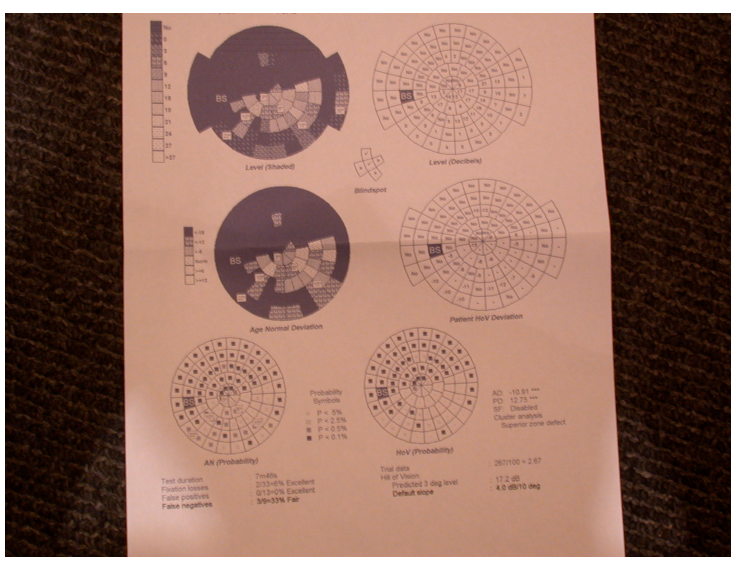
Automated perimetry showing left superior altitudinal field defect. Right eye field is normal (top left), left eye shows dense superior field loss (top right) corresponding to inferior retinal infarction
Fundus Photography - Right Eye: Normal right fundus with healthy optic disc appearance, normal calibre retinal vessels, and clear macula. No signs of vascular occlusion, haemorrhage, or emboli.
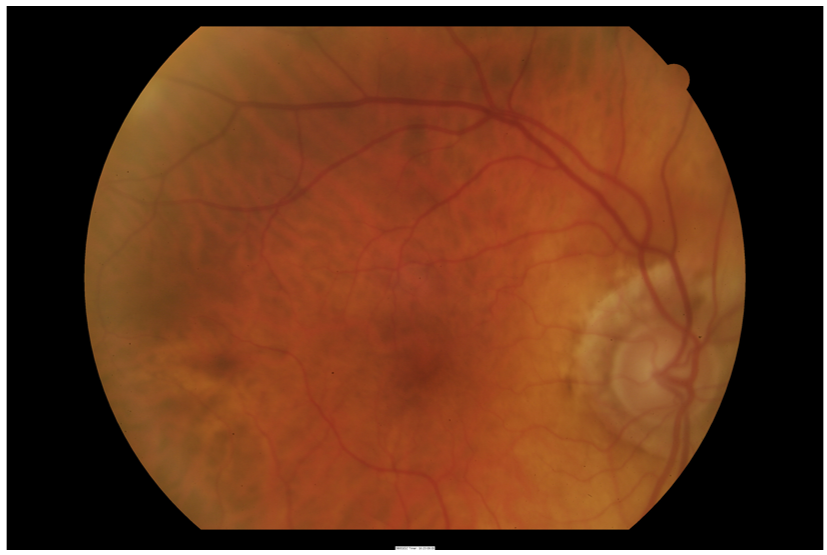
Right fundus photograph showing normal optic disc, retinal vessels, and macula with no pathology
Fundus Photography - Left Eye: Left fundus demonstrates signs of branch retinal artery occlusion affecting the inferior retina. Visible retinal whitening (infarction) in the inferior arcade distribution, consistent with acute ischaemia. An embolus is visible at the bifurcation of the central retinal artery. Attenuated arterioles are present in the affected territory. The superior retina and macula appear relatively preserved.
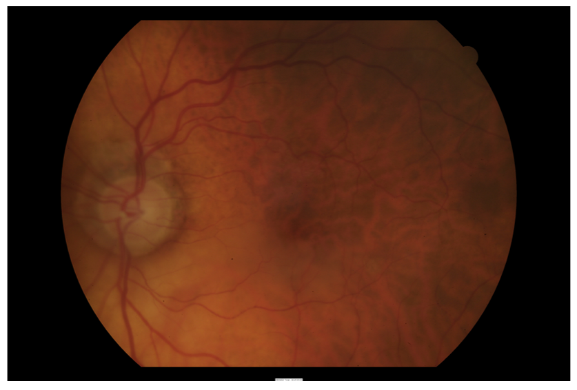
Left fundus photograph showing inferior retinal infarction with retinal whitening, visible embolus at arterial bifurcation, and attenuated inferior arterioles consistent with branch retinal artery occlusion