Quiz: A Lifelong Vision Mystery
Test your understanding of this case with 7 questions.
PATIENT DEMOGRAPHICS
54-year-old male
PRESENTING COMPLAINT
Long-standing bilateral reduced vision first noted during school screening at age 8. Vision has remained relatively stable at 6/12 in both eyes with very gradual decline over the decades. No acute visual symptoms, pain, or photophobia. Difficulty with detailed work and reading small print.
EXAMINATION FINDINGS
Visual Acuity: OD: 6/12, OS: 6/12
Pupils: Equal, reactive, no RAPD
Fundus: Bilateral pale optic discs with temporal pallor more pronounced than nasal. Cup-disc ratio 0.3 bilaterally. Retinal vessels appear normal caliber
INVESTIGATIONS
Fundus Photography: Bilateral pale optic discs with temporal pallor more pronounced than nasal. The optic disc margins are well-defined with cupping consistent with optic atrophy.
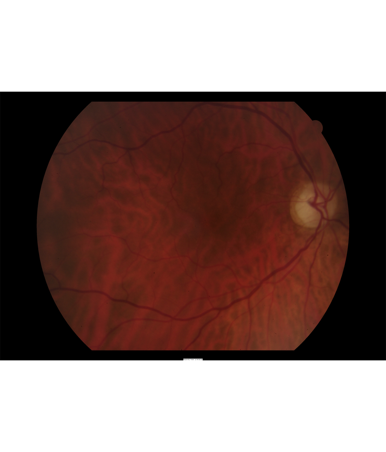
Right eye fundus showing temporal optic disc pallor characteristic of optic atrophy
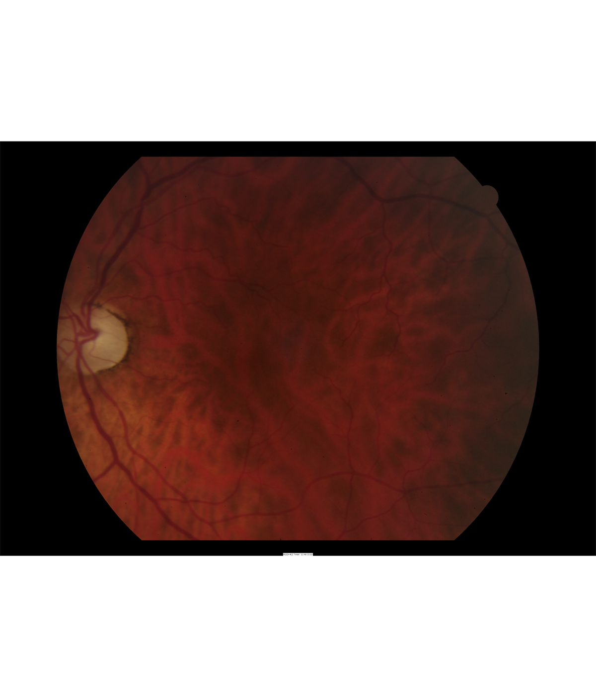
Left eye fundus demonstrating bilateral optic atrophy with similar temporal pallor
Visual Field Test (Humphrey 24-2): Bilateral central and cecocentral scotomas with preserved peripheral fields. Pattern consistent with optic neuropathy.
OCT Optic Nerve: Bilateral thinning of retinal nerve fiber layer, most pronounced in the temporal quadrants. Average RNFL thickness: OD 68μm, OS 65μm (normal >85μm).
Genetic Testing: Heterozygous pathogenic variant in OPA1 gene (c.2713C>T), confirming autosomal dominant optic atrophy.
Family Screening: One of three children carries the same OPA1 mutation but currently has normal vision (age 22). Two children are unaffected.