Quiz: The Stressed Coder's Blood Pressure Crisis
Test your understanding of this case with 8 questions.
PATIENT DEMOGRAPHICS
30-year-old male
PRESENTING COMPLAINT
Blurring of vision in the right eye, headaches, and loss of energy. Recent stress following father's death from motor neurone disease.
EXAMINATION FINDINGS
Visual Acuity: OD: 6/7.5, OS: 6/6
Pupils: Equal, reactive, no RAPD
Fundus: OD: Optic disc oedema with blurred margins, cotton wool spots, flame-shaped hemorrhages. OS: Comparatively normal appearance
INVESTIGATIONS
Fundus Photography: Right eye shows optic disc swelling with blurred disc margins, cotton wool spots, and flame-shaped hemorrhages in the posterior pole. Left eye demonstrates comparatively normal optic disc appearance with minimal retinal changes.
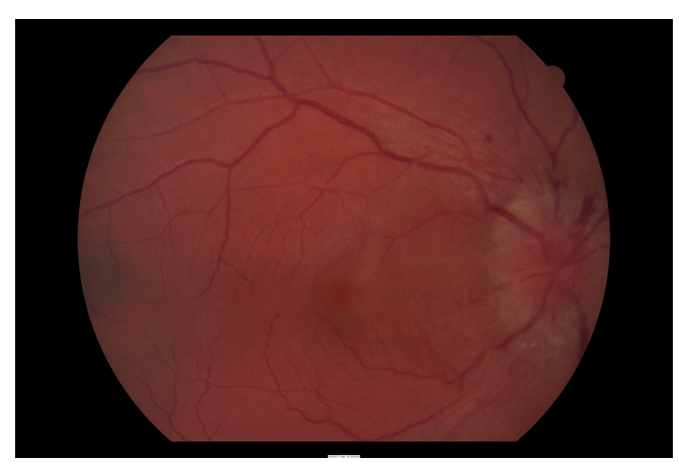
Right posterior pole showing cotton wool spots and retinal hemorrhages consistent with hypertensive retinopathy
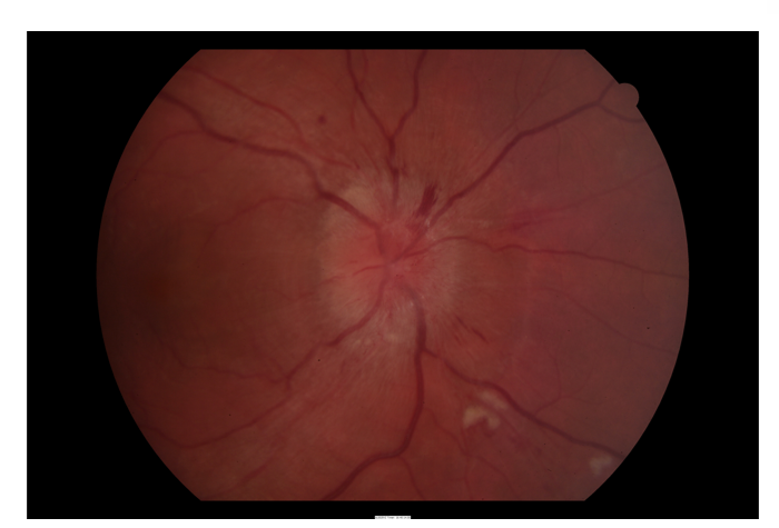
Right optic disc demonstrating significant swelling with blurred disc margins
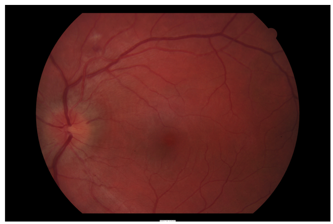
Left posterior pole showing relatively normal retinal appearance
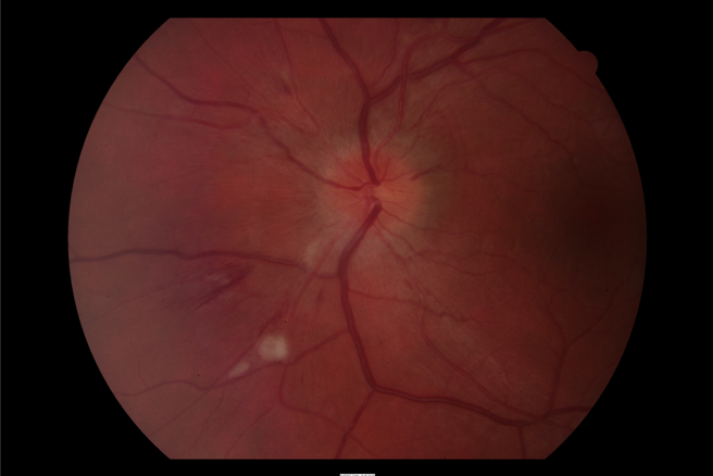
Left optic disc showing minimal changes compared to the right eye
OCT Optic Disc and Macula: Right eye shows significant optic disc swelling with peripapillary fluid and retinal nerve fiber layer thickening. Macular OCT reveals mild retinal thickening. Left eye shows minimal optic disc changes with normal macular architecture, confirming the asymmetric nature of the hypertensive damage.
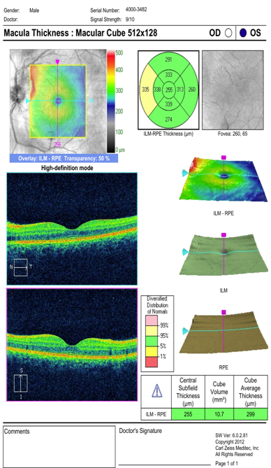
OCT left macula showing normal retinal architecture
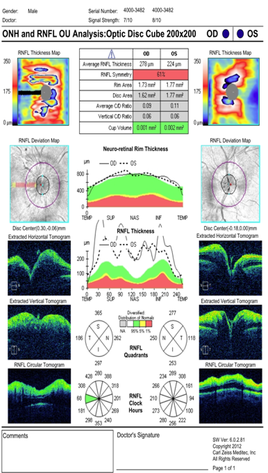
OCT left optic disc demonstrating minimal changes with preserved nerve fiber layer
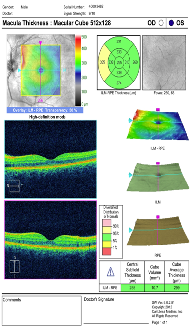
OCT right optic disc showing significant swelling and nerve fiber layer thickening
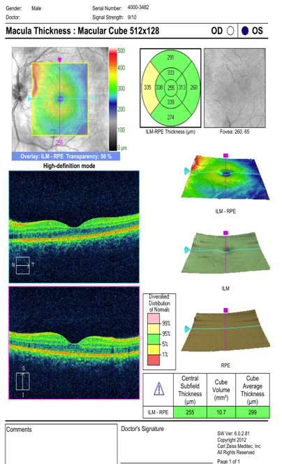
OCT right macula revealing mild retinal thickening secondary to hypertensive changes
Blood Pressure Measurement: 220/145 mmHg - Consistent with hypertensive emergency. Repeat measurements confirmed sustained severe elevation.
Renal Function and Electrolytes: Mild elevation in creatinine suggesting early kidney involvement. No significant electrolyte abnormalities.
Echocardiogram: Left ventricular hypertrophy consistent with longstanding hypertension. Normal systolic function. No structural abnormalities.
Renal Artery Doppler: No evidence of renal artery stenosis. Normal flow patterns bilaterally.
24-hour Urine Catecholamines: Normal levels, excluding phaeochromocytoma as a secondary cause of hypertension.