Quiz: The White Feathery Mystery
Test your understanding of this case with 9 questions.
PATIENT DEMOGRAPHICS
68-year-old male
PRESENTING COMPLAINT
Episodes of transient visual loss in the right eye. Patient reports occasional brief episodes where vision becomes unclear, lasting a few seconds to minutes before returning to normal.
EXAMINATION FINDINGS
Visual Acuity: OD: 6/6, OS: 6/6
Pupils: Equal, reactive, no RAPD
Fundus: OD: White feathery streaks extending from optic disc margin into retina, consistent with medullated nerve fibres. Retinal vessels clearly visible coursing through lesions. OS: Similar but less extensive changes
INVESTIGATIONS
Fundus Photography: Bilateral medullated nerve fibres visible as white feathery streaks radiating from the optic disc margins. Right eye shows more extensive involvement than left eye. Retinal vessels are clearly visible coursing through the white lesions, which is characteristic of this benign congenital anomaly.
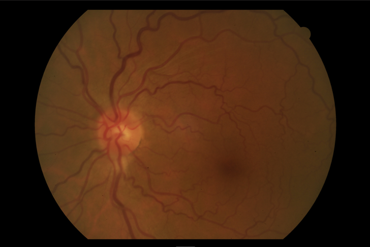
Left fundus showing white feathery streaks of myelinated nerve fibers extending from the optic disc with less extensive involvement
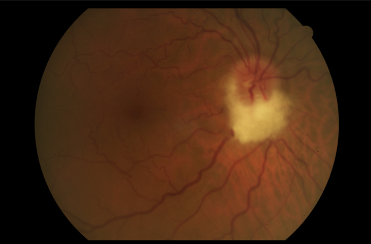
Right fundus demonstrating more extensive white feathery myelinated nerve fibers radiating from the optic disc margin
OCT Retinal Nerve Fiber Layer: Hyperreflective thickened nerve fiber layer in affected areas corresponding to the medullated fibers seen clinically. Normal retinal architecture in unaffected areas.
Visual Field Test (Humphrey 24-2): Bilateral enlarged blind spots corresponding to the areas of medullated nerve fibers. No other field defects detected. Peripheral fields remain intact.
Fundus Autofluorescence: Areas of medullated nerve fibers show hypoautofluorescence, confirming the presence of myelinated tissue anterior to the lamina cribrosa.