Quiz: One Pale Disc, One Swollen Disc
Test your understanding of this case with 9 questions.
PATIENT DEMOGRAPHICS
41-year-old female
PRESENTING COMPLAINT
Visual disturbance with poor vision in one eye and preserved vision in the other. Patient reports difficulty with peripheral vision and occasional headaches.
EXAMINATION FINDINGS
Visual Acuity: OD: 6/60, OS: 6/9
Pupils: Assessment pending - detailed pupil examination required
Fundus: OD: Pale optic disc consistent with optic atrophy. OS: Swollen optic disc with blurred margins consistent with papilloedema
INVESTIGATIONS
Fundus Photography: Right eye shows optic atrophy with pale disc appearance. Left eye demonstrates papilloedema with disc swelling and blurred margins. This combination suggests Foster Kennedy pattern - optic atrophy in one eye with papilloedema in the fellow eye.
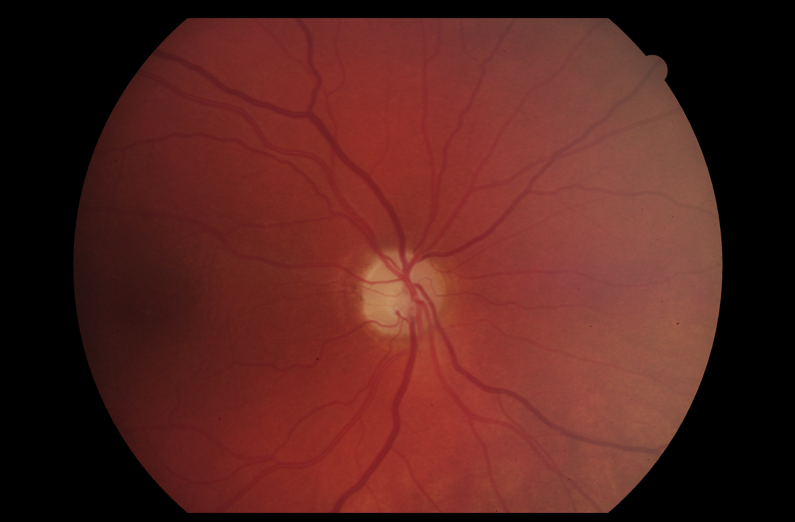
Right eye fundus: optic atrophy with pale disc appearance
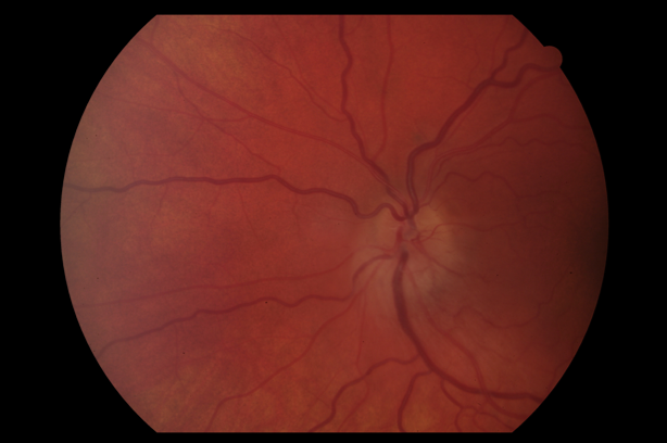
Left eye fundus: papilloedema with disc swelling and blurred margins
OCT Optic Disc Analysis: OCT demonstrates asymmetric retinal nerve fiber layer changes: right eye shows significant RNFL thinning consistent with optic atrophy, while left eye shows RNFL thickening consistent with papilloedema. This confirms the clinical findings and supports the diagnosis of Foster Kennedy pattern.
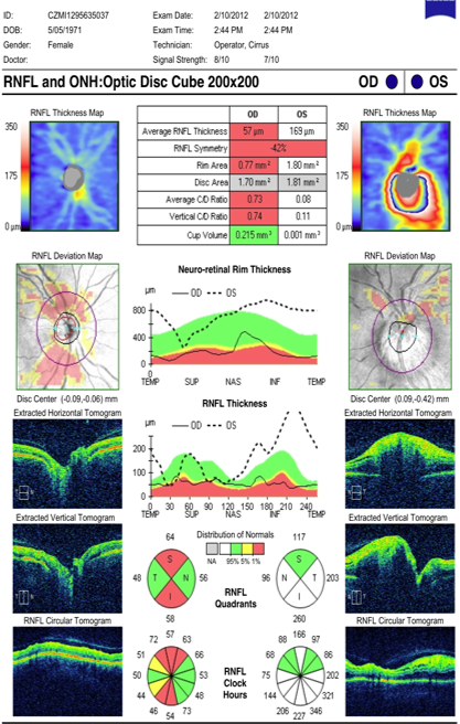
OCT: asymmetric RNFL - right eye thinning (atrophy), left eye thickening (swelling)
Visual Field Assessment: Right eye: Superior altitudinal defect consistent with optic nerve damage. Left eye: Enlarged blind spot secondary to papilloedema with otherwise preserved peripheral fields.
Neuroimaging (MRI Brain and Orbits): Pending urgent imaging to exclude intracranial space-occupying lesion compressing optic pathways and causing raised intracranial pressure.