Quiz: The Cardiac Patient's Vanishing Vision
Test your understanding of this case with 11 questions.
PATIENT DEMOGRAPHICS
44-year-old male
PRESENTING COMPLAINT
Sudden-onset visual field loss. Patient describes loss of vision in part of the visual field. No eye pain or headache reported. The patient was found to have viral cardiomyopathy on echocardiography performed as part of the stroke workup. Visual acuity was preserved bilaterally at 6/6. Pupils were equal and reactive with no relative afferent pupillary defect.
EXAMINATION FINDINGS
Visual Acuity: OD: 6/6, OS: 6/6
Pupils: Equal and reactive, no relative afferent pupillary defect (RAPD)
Fundus: Fundus examination unremarkable bilaterally
INVESTIGATIONS
Visual Field Testing (Automated Perimetry): Dense left homonymous hemianopia with subtle right homonymous inferior quadrantic loss. The combination of dense hemianopia on one side with subtle contralateral defect suggests bilateral occipital involvement.
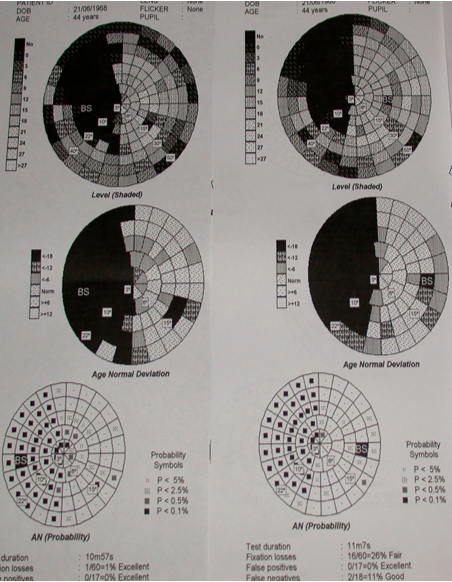
Automated perimetry showing dense left homonymous hemianopia and subtle right homonymous inferior quadrantic loss
MRI Brain: Demonstrates bilateral occipital cortex infarcts, confirming the clinical localization from visual field testing.
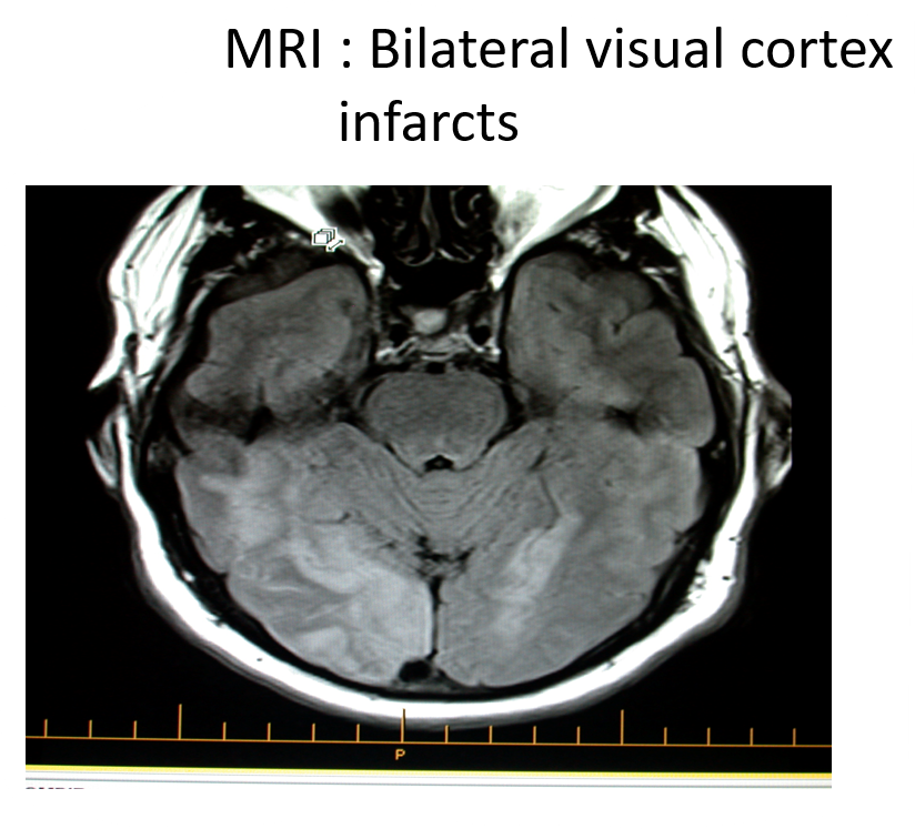
MRI Brain: Bilateral visual cortex infarcts in occipital lobes
Echocardiography: Viral cardiomyopathy with impaired ventricular function identified as a high-risk source for intracardiac thrombus formation and systemic embolism.