Quiz: The Painful Eye That Won't Move
Test your understanding of this case with 16 questions.
PATIENT DEMOGRAPHICS
84-year-old female
PRESENTING COMPLAINT
Progressive double vision (both vertical and horizontal) and deep left orbital pain over several weeks.
EXAMINATION FINDINGS
Visual Acuity: Visual acuity testing was limited by ptosis and diplopia but was estimated at approximately 6/9 bilaterally. Formal documentation was incomplete at initial presentation.
Pupils: Left pupil involvement suspected (CN III palsy with possible pupillary involvement)
Fundus: No papilloedema or retinal abnormalities noted
INVESTIGATIONS
Clinical Photographs - Ptosis and Eye Position: Frontal view demonstrates left upper lid ptosis. Close-up examination shows the left eye in a position consistent with CN III palsy (eye down and out position).
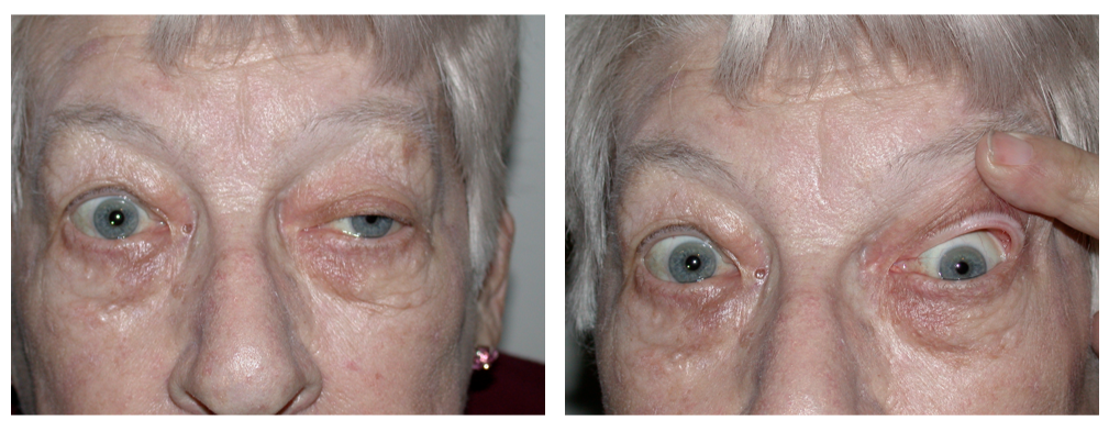
Clinical photographs showing left ptosis and abnormal eye position at rest
Extraocular Movement Assessment: Systematic assessment of eye movements using fixation targets demonstrates severe restriction in multiple directions of gaze. Left eye shows marked limitation in elevation, depression, and adduction (CN III palsy) with impaired abduction (CN VI involvement). Multiple positions of gaze documented showing the extent of ophthalmoplegia.
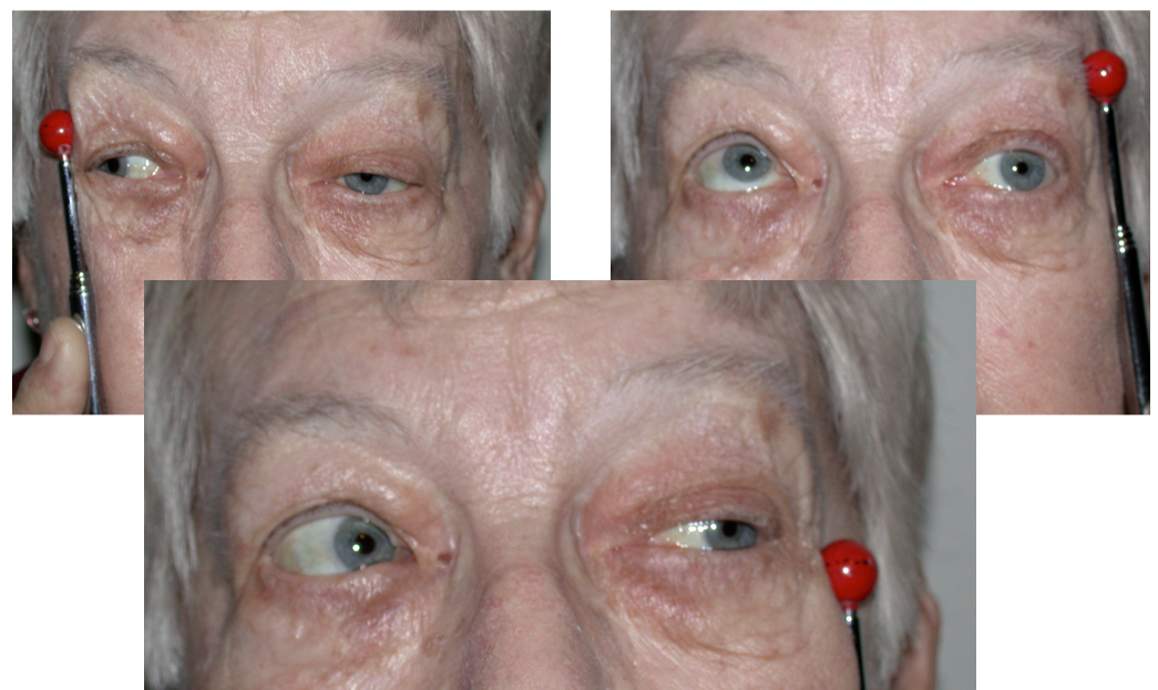
Extraocular movement testing with fixation targets demonstrating left CN III and CN VI palsies with restricted motility in all directions
CT Brain (Axial Views - Non-contrast and Contrast Comparison): Axial CT imaging in both non-contrast and contrast-enhanced phases reveals a cavernous sinus lesion consistent with an internal carotid artery aneurysm. The contrast-enhanced images show characteristic filling of the aneurysmal sac, confirming the vascular nature of the lesion. The location within the cavernous sinus explains the multiple cranial nerve palsies.
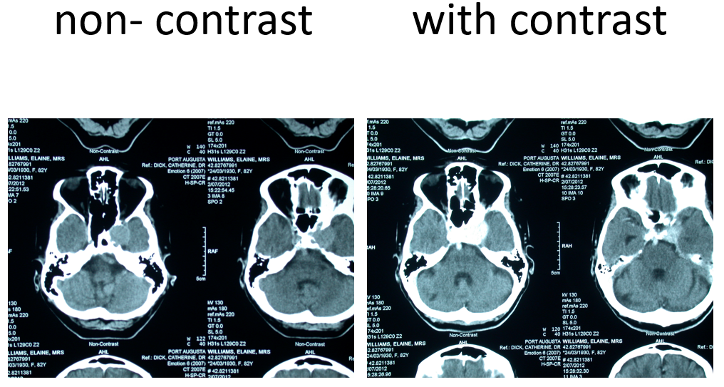
CT brain comparison: Non-contrast (left) and contrast-enhanced (right) axial views demonstrating left cavernous sinus internal carotid artery aneurysm with contrast filling
Neurological Examination: Pain distribution corresponds to left ophthalmic division (V1) of trigeminal nerve. Features suggestive of Horner's syndrome (miosis, mild ptosis) from sympathetic fibre involvement.